 Organic Workflow
 Peptide Workflow
 Scale-Up Flash Purification
  Sample Preparation
 Biomolecule Purification
 Oligo synthesis
 Scavengers and Reagents
 Service & Support
 Accessories & Spare parts
 Investors
 Reports & News
 The Share
 Corporate Governance
 Calendar
 Sustainability
 Our Offering
 Our History
 Our Locations
 Leadership
Now that you have your purified plasmid, you will need to get an idea of its quality. Transfection efficiency is highly influenced by the quality of the plasmid, so it’s important to get a full picture. There are a few factors to plasmid quality, and you will want to look at all of them to make sure your samples will be successful in your downstream processing. Here, I will explain how to measure the absorbance ratios of your sample, successfully read an agarose gel, and the importance of low endotoxin values.
Measure absorbance ratios
The first step after purifying plasmid is always to quantify your yield, using a NanoDrop™ or a similar system. A NanoDrop™ reads concentration by assessing the absorbance at 260 nm, this is the wavelength at which DNA is absorbed. The NanoDrop™ will also report additional absorbance values, that give you some insight into the purity of your plasmid, and any possible contaminants your sample may contain. There are two industry-standard absorbance value ratios, A260/A280, and A230/A260. The absorbance of DNA is compared to the absorbance of contaminants, and the ratio presented gives you insight into how much DNA is in your samples compared to possible contaminants.
The A260/A280 ratio is used to measure DNA purity. A ratio of around 1.8 is generally accepted as “pure.” If the ratio is lower (<1.6), the sample may contain proteins, which absorb strongly at or near 280 nm. This ratio is affected by the solvent used, for example, acidic solutions will underrepresent the ratio by 0.2-0.3, and a basic solution will overrepresent the sample by the same amount. Therefore, it is important to equalize the pH of your samples before comparing their A260/A280 ratios or to blank with the correct buffer. Generally, the elution buffer is ideal for blanking so it can account for any components of the elution buffer.
The A230/A260 ratio is used as a secondary measure of DNA purification. A ratio between 2.0 and 2.2 is considered “pure.” If the ratio is lower, the sample may contain phenols, guanidine GCL, EDTA, carbohydrates, lipids, or salts, which are all things that absorb at 230 nm. However, this ratio is easily altered when a saline elution buffer is used to dissolve the DNA, so it is important to consider that a higher ratio may still be considered pure when eluting in salt.
Run a gel electrophoresis
To check the quality of your plasmid DNA, you can run a gel. A gel will tell you a couple of things about your plasmid. First, it will tell you the plasmid’s confirmation: supercoiled, linear, or circular. It will also tell you if there is any genomic DNA or RNA contamination in your sample. Finally, it will tell you if you over-lysed or denatured your sample.
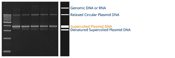
Transfection will be most efficient with supercoiled plasmids, so you are looking for the band labeled “supercoiled plasmid” in orange on the image above. If your sample is slightly over-lysed or nicked, you may see a denatured supercoiled plasmid band, slightly below where the supercoiled plasmid band would be. A denatured supercoiled plasmid will be single-stranded, so it will be about half the mass of your supercoiled plasmids, therefore it will travel farther down the gel. If you see a genomic DNA band at the top, or ribosomal RNA bands at the bottom, your alkaline lysis may not be efficient. Alkaline lysis causes genomic DNA to precipitate out of the sample, so it cannot reanneal after the lysis step. You may need to separate the supernatant more fully from the cell debris or optimize your alkaline lysis step in order to remove genomic DNA more effectively. Additionally, your RNase might not be effective if you see ribosomal RNA bands.
Measure Endotoxin
An endotoxin, also known as a lipopolysaccharide or LPS, is a pyrogen (fever-producing compound) that is found in the cell membrane of Gram-negative bacteria, such as E. coli. Aside from its fever-producing capabilities, which make it dangerous when found in drugs and medical devices, it also forms micelle structures that mimic DNA in size and charge. As a result, endotoxin micelles compete with DNA as transfection reagents, decreasing the efficiency of your transfection. High-quality plasmid DNA needs to have low or no endotoxin.
Endotoxin is measured using LAL (Limulus amebocyte lysate), an aqueous extract of blood cells from Atlantic horseshoe crabs. LAL reacts with endotoxin, coagulating, giving scientists the ability to measure the presence of endotoxin in their samples. Charles River created a Bacterial Endotoxins Test (BET) that is readily available to test samples
To learn more about how to automate your plasmid prep process and obtain reliable transfection-grade DNA, click on the button below.